
Products

Description:
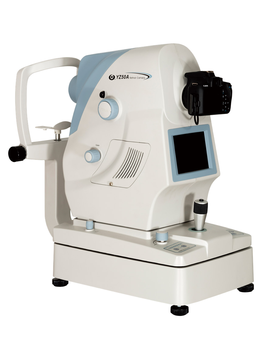
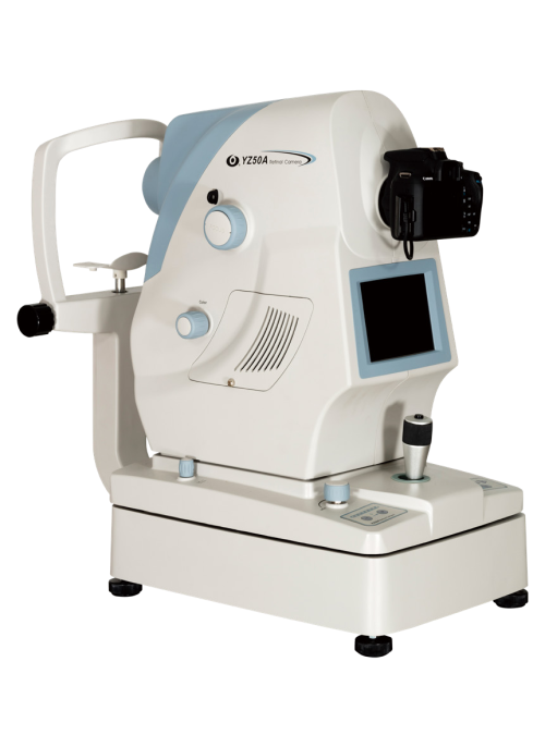
The YZ50A Serious Non-Mydriatic Fundus Camera is used to observe and photograph the posterior segment of the eye, including the retina, macula, and posterior pole. The instrument uses infrared light as the illumination source and features a focus split indicator to show the amount of focus deviation on the fundus observation screen. It also locates the accurate position using pupil positioning spots. The instrument is easy to operate, flexible, and reliable for medical use.
|
Specifications |
|
|
Eyepiece magnification |
12.5× |
|
Large objective lens focal length |
F=215 mm |
|
Working distance |
170mm |
|
Total magnification of main microscope |
4.6× ~ 27× motorized and manual control continuous magnification |
|
Field diameter |
4.6×(Φ46mm) 27×(Φ8.5mm) |
|
Secondary microscope magnification |
6×、10×、14× |
|
Adjustment Diopter |
+5D ~ - 5D |
|
Adjustment Range of Pupil Distance |
55mm ~ 75mm (main microscope is 45mm ~ 80mm) |
|
Illumination Source |
12V/100W,cold reflection halogen lamp for medical use |
|
Lighting mode |
6 ° cold light source coaxial lighting; 26 ° oblique lighting |
|
Lighting range |
Φ45(F=215mm) |
|
Tiny focus stroke |
≥40mm |
|
Fine focusing speed |
≤2mm/s |
|
X-Y movement range |
50mm × 50mm (X - ← 0 ~ - 25; in situ 0; X+→ 0 ~ +25; Y- ↓ 0 ~ -25;Home position 0; Y+ ↑ 0 ~ +25) |
|
X-Y moving speed |
≤2mm/s |
|
Vertical adjustment range |
880mm ~ 1420mm |
|
Reaching Radius of Arm |
1230mm |
|
Input power |
170VA |
|
Power Supply |
AC110V/220V±10%,50Hz/60Hz |

